NMR Metabolomics in Animal Models: A Key Tool for Translational Research

In biomedical research, animal models represent a fundamental tool for understanding the pathophysiological mechanisms of human diseases and for exploring new therapeutic strategies. They allow for the controlled reproduction of complex clinical conditions, the study of disease progression—particularly in chronic or metabolic disorders—and the evaluation of interventions before their application in humans.

In this context, metabolomics, particularly that based on nuclear magnetic resonance (NMR), has emerged as an essential technique for globally and quantitatively characterizing metabolic profiles in living organisms. NMR enables the detection and quantification of dozens of metabolites in a single sample, without requiring chemical derivatization or destruction, offering high reproducibility and comparability between studies. Its application to biological matrices such as serum, plasma, urine, or tissue provides a precise “metabolic fingerprint,” which is key for identifying functional biomarkers, biochemical alterations, or treatment responses in murine, porcine, or even large-animal models.

At Biosfer Teslab, research drives our mission. We actively collaborate with academic and clinical research groups that use animal models to study metabolic, cardiovascular, hepatic, and endocrine diseases, offering advanced tools for molecular characterization. Thanks to our experience in NMR-based metabolomics, our analyses contribute to a deeper and more functional understanding of the biological processes involved in each model. This integration not only helps identify disease biomarkers and mechanisms but also supports the validation of therapeutic interventions with scientific rigor and translational potential.

What Can NMR Metabolomics Contribute to Animal Model Research?

Metabolomics allows for the global and quantitative analysis of small metabolites present in biological fluids and tissues, offering a functional snapshot of an organism’s metabolic state. Applied to animal models, this discipline is a powerful tool to investigate the pathophysiology of complex diseases and evaluate therapeutic interventions under controlled conditions.

One of the strengths of metabolomics is its ability to provide a functional and systemic view of metabolism, reflecting the biochemical state of the organism at a specific moment. In animal models, this approach enables the detection of metabolic patterns associated with disease processes and the generation of hypotheses about underlying mechanisms in disease development or progression. It complements other molecular techniques and is useful for biomarker discovery, experimental intervention assessment, and understanding biological responses to experimental conditions or treatments.

Advantages of Using NMR-Based Metabolomics in Animal Models

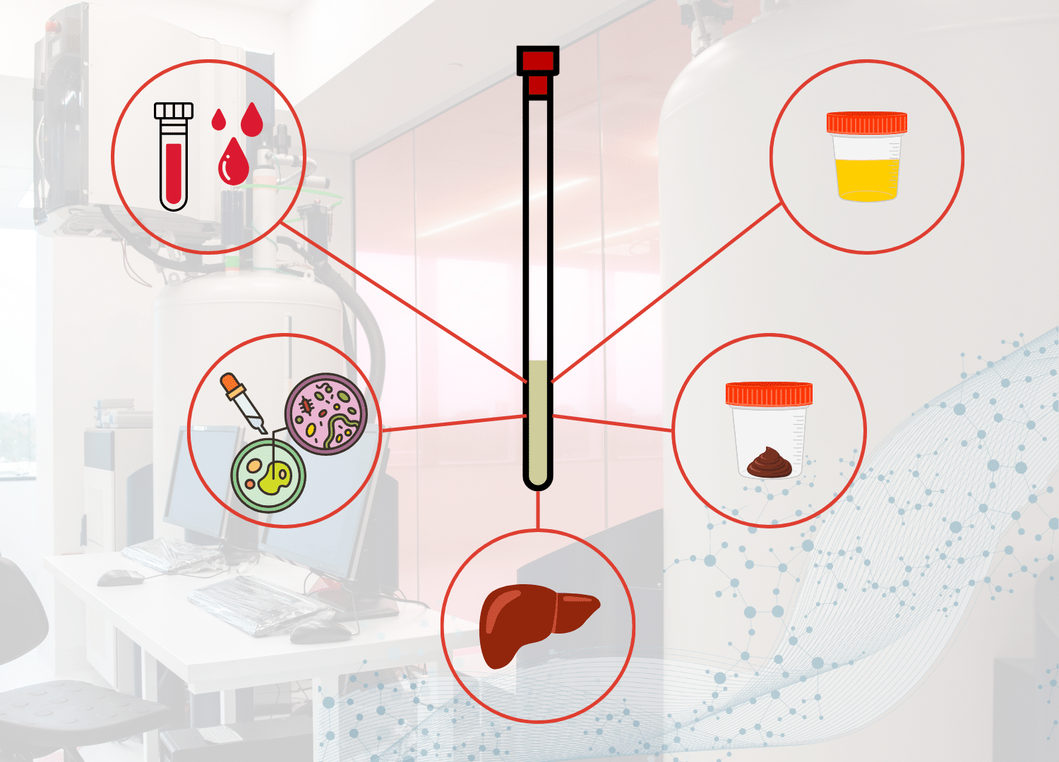
Nuclear Magnetic Resonance (NMR) applied to metabolomics offers several advantages that make it particularly suitable for preclinical studies involving animal models:

  • Non-destructive and low-preparation: Samples are analyzed in their original state, without derivatization, preserving them for future use and maintaining metabolite integrity.
  • High reproducibility and analytical robustness: NMR provides highly stable and comparable results across experiments, institutions, and time points—ideal for longitudinal and multicenter studies.
  • Suitable for diverse biological matrices: Works across serum, plasma, urine, feces, and tissues using standardized protocols, ensuring directly comparable outputs.
  • Ideal for small sample volumes: Especially in models like mice or rats where sample quantities are limited, NMR yields rich functional data from as little as 50 µL.

NMR-sample-analysis.jpg

Applications of NMR Metabolomics in Animal Models

NMR Metabolomics for Evaluating  Therapeutic Strategies in NASH

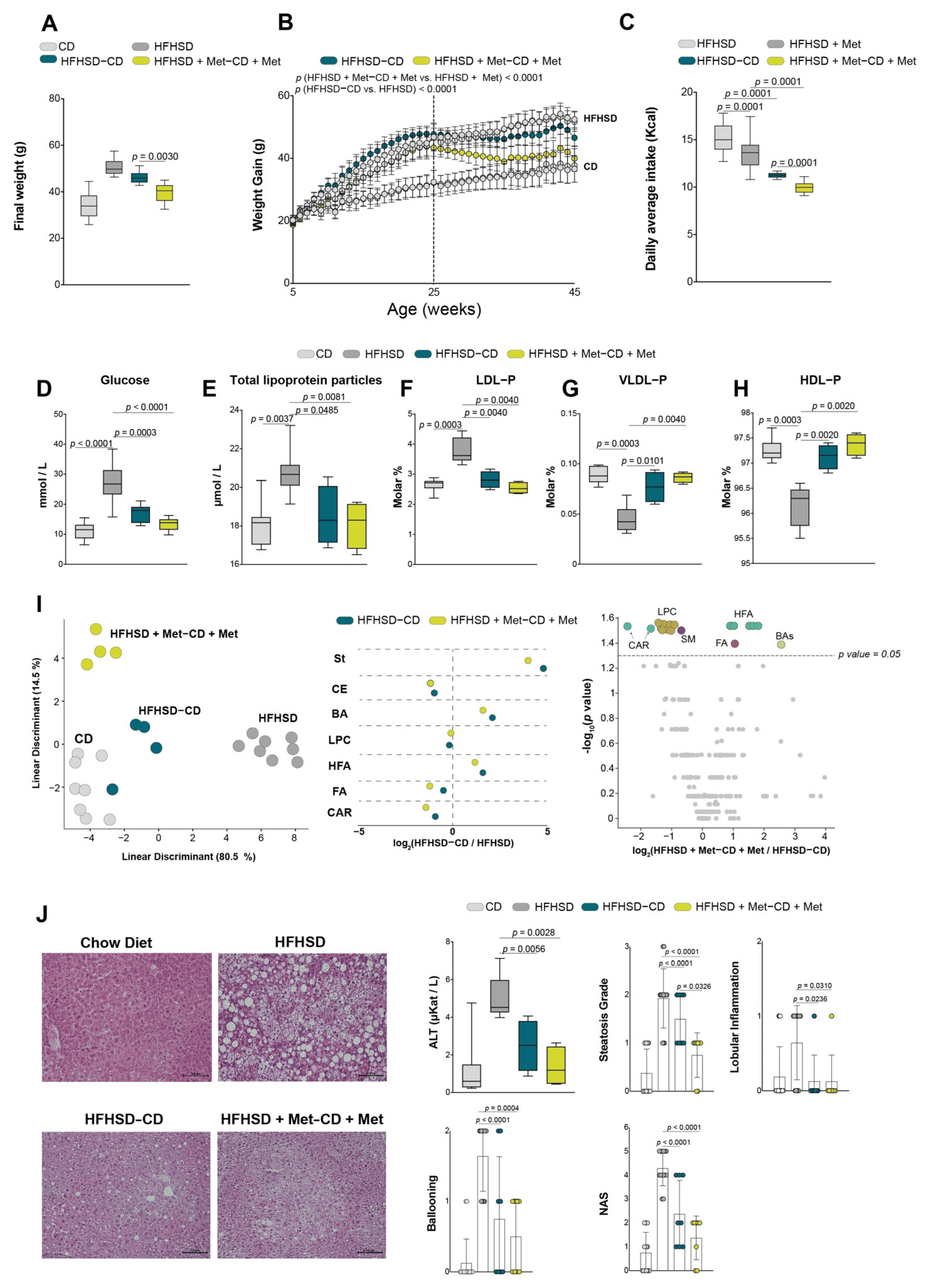
This study, conducted in a murine model of non-alcoholic steatohepatitis (NASH) induced by a high-fat, high-sucrose diet, evaluated the effect of metformin administered alone or in combination with dietary intervention. Metformin alone did not prevent NASH development or improve lipid and histological parameters. However, when combined with dietary changes, it showed synergistic effects: greater weight reduction and more significant reversal of hepatic steatosis. To assess these effects, the study incorporated NMR-based lipidomic metabolomics (Liposcale®) along with semi-targeted mass spectrometry analysis. These technologies revealed specific remodeling in hepatic and adipose tissue metabolism, such as increased polyunsaturated fatty acids (PUFA) and reduced cholesterol esters (CE), confirming the metabolic impact of the combined treatment.

Reference:
Baiges-Gaya G, Rodríguez-Tomàs E, Castañé H, et al. Combining Dietary Intervention with Metformin Treatment Enhances Non-Alcoholic Steatohepatitis Remission in Mice Fed a High-Fat High-Sucrose Diet. Biomolecules. 2022;12(12):1787. doi:10.3390/biom12121787.

Figure 2. Combined effects of dietary intervention and metformin in the NAFLD remission: (A) final body weight; (B) body weight curve; (C) caloric intake (n = 8/group). The concentration of (D) glucose (n = 8/group); (E) total lipoprotein parameters (n = 4/group); (F–H) VLDL-p, LDL-p, and HDL-p molar abundance (expressed as a percentage of total measured lipoprotein particles) (n = 4/group); (I) lipid signature analysis and significant alterations in liver comparing the effects of calorie restriction with or without metformin versus obese mice lipidome (n = 4/group); (J) representative histology images of liver sections with the NAFLD activity score, and alanine aminotransferase activity (n = 8/group). Scatter and bar plots are presented as means and SD, while box plots are shown as means, maximum and minimum. p values < 0.05 are considered significant. (Wilcoxon-rank sum test). ALT: alanine aminotransferase; BA: bile acids; CAR: carnitines; CD: chow diet; CE: cholesterol esters; FA: fatty acids; HDL: high-density lipoproteins; HFA: hydroxy fatty acids; HFHSD: high-fat high-sucrose diet; LDL: low-density lipoproteins; LPC: lysophosphatidylcholines; Met: metformin; NAS: NAFLD activity score; p: particles; SM: sphingomyelins; St: steroid hormones; VLDL: very low-density lipoproteins

Fecal NMR Metabolomics: Early Biomarkers for Gut Health in Animal Models

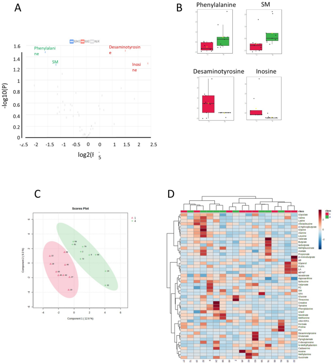
This study, published in Scientific Reports (2025), evaluated the impact of neonatal management on gut health in male dairy calves using fecal metabolomics by NMR. A controlled experimental design involved three groups of Holstein calves exposed to different colostrum intake, transportation, and feeding strategies. The most nutritionally restricted group (LCRS) showed significantly elevated fecal lactoferrin levels, indicating gut inflammation. The NMR metabolic profiling identified over 50 fecal metabolites, highlighting key alterations such as reduced butyrate (a crucial short-chain fatty acid for gut integrity) and elevated levels of stress-related compounds like formate, proline, and creatine. Multivariate PLS-DA analysis showed clear group separation, illustrating how fecal metabolomics can sensitively capture early nutritional and environmental stress effects.

Reference:
Bassols A, Amigó N, Pérez-Rodado M, et al. Fecal metabolomics to understand intestinal dysfunction in male dairy beef calves at arrival to the rearing farm. Sci Rep. 2025;15(1):6887. doi:10.1038/s41598-025-90407-3.

Figure 3.Metabolomic analysis considering the three groups CTR, LCMR and LCRS. (A) Metabolic profiles of the CTR (group 1, red), LCMR (group 4, green) and LCRS (group 5, blue) fecal samples using PLS-DA score plots. Each shape indicates one sample colored according to the group with ellipse indicating the 95% confidence region. (B) Metabolites with VIP > 1.0 corresponding to component 1. The colored boxes on the right indicate the relative concentrations of the corresponding metabolite in each group under study. (C) Hierarchical clustering heatmap from groups CTR (group 1, red), LCMR (group 4, green) and LCRS (group 5, blue).

NMR Metabolomics to Evaluate Nutritional Interventions

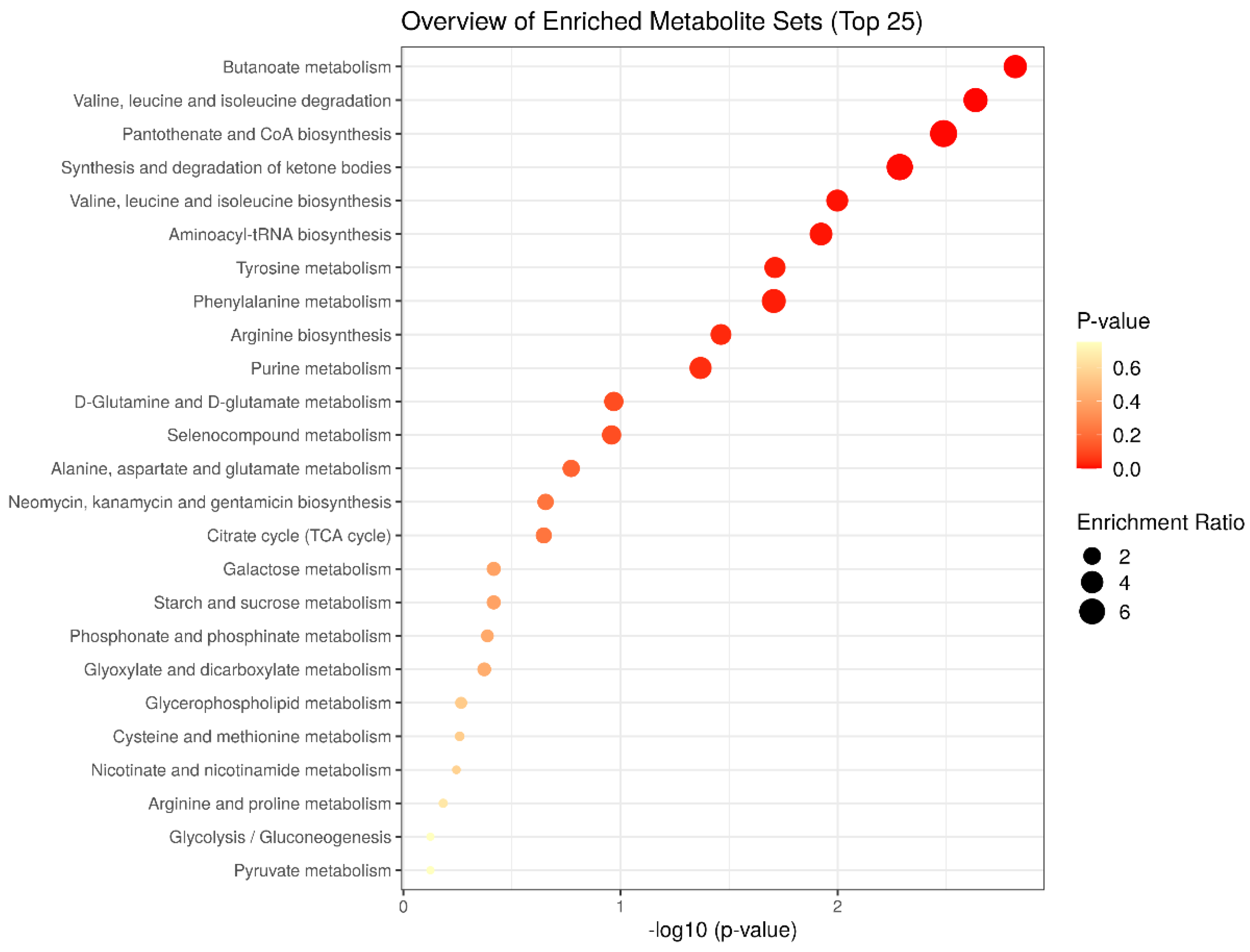
This study, published in Nutrients (2022), examined the metabolic effects of chronic cocoa consumption in a type 2 diabetes animal model (Zucker diabetic rats). Using a non-targeted NMR-based urinary metabolomics approach, researchers identified a distinct metabolic signature linked to cocoa intake, including increased levels of branched-chain amino acids (BCAAs: valine, leucine, isoleucine) and reduced acetoacetate—suggesting improved insulin sensitivity and reduced hepatic gluconeogenesis and ketogenesis. Significant changes were also observed in key metabolic pathways such as butanoate metabolism and CoA biosynthesis. These findings support cocoa’s protective role in diabetes and demonstrate how NMR metabolomics delivers functional insights into the systemic impact of nutritional interventions.

Reference:
Fernández-Millán E, Ramos S, Álvarez-Cilleros D, et al. Urinary Metabolomics Study on the Protective Role of Cocoa in Zucker Diabetic Rats via 1H-NMR-Based Approach. Nutrients. 2022;14(19):4127. https://doi.org/10.3390/nu14194127

Figure 4. Metabolite set enrichment analysis of significantly altered urine metabolites according to the KEGG database.

Do You Work with Animal Models?

Ready to take your research further?

Discover how NMR-based solutions can support your next study

Contact our team to start the conversation.

Practical details: Biosfer Teslab Portfolio